Reading Time: 9 minutes

INTRODUCTION

Development of nano-theranostic platforms, particularly those based on quantum dots (QDs) for simultaneous sensing, imaging and therapy, have evinced interest in researchers in recent years. The major advantage with the use of QDs is the possibility of achieving high precision in controlling the conductive properties of the material through the control of their crystal size.

The physicochemical properties of QDs (Figure 1) have enabled their application in various domains such as single-electron transistors, solar cells, LEDs, displays, lasers, photodetector devices, photocatalysts, photovoltaic devices, quantum computing, forensic, microscopy, and medicine.[1]

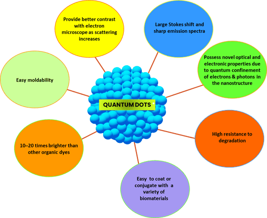
physicochemical-properties-of-quantum-dots

Figure 1. Physicochemical properties of Quantum Dots

Though QDs have many advantages, there are significant limitations in their widespread use. Major challenges include low biological specificity, low aqueous solubility, complicated surface chemistry, poorly controlled biodistribution to target tissues, and potential long-term toxicity.[2] Efforts are in progress to improve long-term stability in biological buffers, quantum yield following bioconjugation, and clearance from the body.

QUANTUM DOTS IN MEDICINE – RECENT TECHNOLOGIES

The unique optical properties of QDs have led to their use in the biomedical field for imaging/ tracking, diagnostics, drug delivery, tissue engineering, cancer therapy, multicolor optical coding, protein detection, as sensors/ biomarkers/ nanoprobes, antimicrobial agents and transgenic vectors, and single molecule probes, among many other applications.[3,4]

Some of the major applications of quantum dots are presented below.

Imaging & Diagnosis:

QDs are extensively used as in vivo imaging agents. Specific antibodies coupled to near-IR (NIR) QDs with a polymer coating have been frequently used for tumor-targeted imaging.[4] Researchers have attempted to create in vitro and in vivo testing methods for imaging nanoscale and microscale structures by cutting down on the quantity of Cd released in the cells. Recent studies have focused on the development of functionalized QDs so as to create diagnostic tools that enable more sensitive ex-vivo identification of disease-relevant biomolecules.[2,4]

The poor transmission of visible light through the biological tissue using fluorescence imaging, has prompted researchers to consider the possibility of using NIR optical window (700–1,700nm) for conducting deep-tissue optical imaging using QDs.

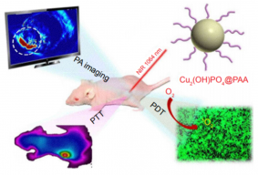
Use of QDs in image-guided therapy has particularly been considered to be effective with high precision for the treatment of cancer. One such development is the fabrication of polyacrylic acid-coated Cu2(OH)PO4 quantum dots (Cu2(OH)PO4 @PAA QDs) that exhibit a strong NIR photo-absorptive ability as a “three-in-one” multipurpose cancer theranostic system (Figure 2). This has been used in a combination of photodynamic therapy (PDT) and photothermal therapy (PTT) coupled with photoacoustic imaging modality. In the process of NIR excitation, a local hyperthermia and reactive oxygen species (ROS) were generated, recognizing the synergetic PTT/PDT therapeutic effects. These QDs have been found to be good candidates for in vivo photoacoustic imaging of the tumor site for realizing an imaging-guided therapeutic process.[5,6]

proposed-cu2ohpo4paa-three-in-one-multifunctional-theranostic-platform

Figure 2. Proposed Cu2(OH)PO4@PAA “three-in-one” multifunctional theranostic platform[6]

Researchers from Zhongnan Hospital, Wuhan University introduced an effective photothermal agent by packaging black phosphorus quantum dots (BPQDs) into exosome vector (EXO) through electroporation. The resulting BPQDs@EXO nanospheres (BEs) exhibit good biocompatibility, long circulation time, excellent tumor targeting ability, and highly efficient tumor ablation in vivo.[7]

Colloidal Quantum Dots (CQD) have evinced considerable attention from researchers for application in biomedical diagnosis, imaging, biochemical analysis and stem cell tracking. Quasi core/shell lead sulfide/reduced graphene oxide CQDs with NIR emission (1100 nm) have been developed for potential bioimaging applications. The graphene dot-based heterostructure semiconductor particles possessing several surface functional groups, particularly the presence of carboxylate and epoxy groups on the surface of hybrid nanocrystals, render good water dispersibility and stability that are essential for biomedical applications.[8]

Bioimaging probes incorporating QDs have found applications in the identification and monitoring of organelles in living cells. Organelle specificity is achieved by functionalizing probe surfaces with chemical groups that can react with antibodies capable of targeting specific organelle-protein epitopes.

Researchers from the University of Massachusetts have developed ZnS-capped CdSe QDs encapsulated within polystyrene (PS) nanocolloids via Pickering miniemulsion using laponite nanoclay platelets as solid-stabilizers. Aminopropyltriethoxysilane (APTES) has been used to modify the surface of the platelets, which are biotinylated by reacting sulfo-NHS-Biotin via the APTES amine group. PS-QD nanocolloids exhibit easy water-dispersibility and long-term photostability, with no cytotoxicity to living cells. These attributes demonstrate the applicability of PS-QDs nanocolloids as long-lived fluorescent bioprobes for in vitro intracellular imaging.[9]

Researchers from the Seoul National University College of Medicine fabricated alloy-typed core/shell CdSeZnS/ZnS quantum dots (alloy QDs) with higher quantum yield and stability during the surface modification for hydrophilization compared to the conventional CdSe/CdS/ZnS multilayer quantum dots (MQDs). Alloy QDs and MQDs, after conjugation with folic acid, were successfully used for targeting human KB cells.[10]

QDs have also found application as vehicles for delivery of photosensitizers in cancer diagnosis and photodynamic therapy (PDT).  Researchers from China have studied pH-responsive fluorescent graphene QDs (pRF-GQDs) for the detection of cancer in the early stages of tumor formation. The high sensitivity against cancerous cells, high safety, and fluorescence switching between healthy weaves and tumors have enabled the application of pRF-GQDs as a prospector. The pRF-GQDs have minimal toxicity, and display a sharp fluorescence transition between green and blue at pH 6.8, matching the acidic extracellular microenvironment in solid tumors. This unique fluorescence switch property helps in differentiating tumors from normal tissues. pRF-GQDs also bear Upconversion photoluminescence (UCPL) property. The combination of UCPL and fluorescence switch enables detection of solid tumors of different origin at an early stage. Hence, pRF-GQDs have great potential as universal probes in fluorescence-guided cancer surgery and cancer diagnosis.[11]

Researchers from Jawaharlal Nehru University have developed cadmium-free water-soluble silver indium sulfide (AgInS2 or AIS) and AgInS@ZnS (or AIS@ZnS) core-shell QDs through a microwave-assisted approach using glutathione (GSH) as stabilizer. The long fluorescence lifetimes (>300 ns), low cytotoxicity, optical and colloidal stability in the physiological pH range of these aqueous dispersible core-shell QDs have enabled therapy as well as monitoring live systems for extended periods of time. Photo-stability and photodynamic efficacy are enhanced due to the shelling of the core QDs, thus exhibiting high potential for photochemotherapy of cancer.[12]

Cryo-imaging has been employed by researchers for labeling cells. Generation of fluorescence for the purpose is achieved through various means such as bioconjugated nanocrystal dyes, lipophilic dyes and cytoplasmic dyes. Researchers from Chiang Mai University, Case Western Reserve University, BioInVision Inc., and Johns Hopkins University, studied the use of multispectral cryo-imaging for stromal cell imaging. The study involved use of cryo-imaging and software to analyze biodistribution of human mesenchymal stromal cells (hMSCs), in mouse models of graft-versus-host-disease (GVHD) following allogeneic bone marrow transplantation (BMT). The researchers employed 1 x 10⁶ human MSCs labeled with red QDs (Qtracker® 625, Life Technologies), for the study.[13]

QDs have also proved to be a possible tool for the identification of the dynamics of specific proteins in living cells. Researchers from Vanderbilt University explored the possibility of using QDs to label neuronal proteins in a single QD imaging format.[14] QD single-particle tracking is a robust technique to target a wide variety of membrane proteins.[15] Phenylboronic acid (PBA)-functionalized graphitic carbon nitride fluorescent QDs (PCQDs) were synthesized and applied to detect sialic acid on the surface of cells and tissues.[16]

Researchers from National Tsing Hua University developed a dual aptamer assay for detection of Acinetobacter baumannii (AB) on an electromagnetically-driven microfluidic platform. This involves an integrated microfluidic device for AB diagnosis utilizing a dual aptamer assay for point-of-care (POC) applications. Magnetic beads coated with AB-specific aptamers are being used to capture bacteria, and QDs being bound to a second aptamer to quantify the number of bacteria with a light-emitting diode (LED)-induced fluorescence module integrated with the device.[17]

Drug delivery:

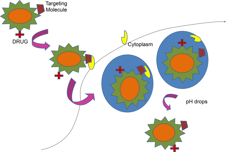
The ability to target the delivery system and easily differentiate unhealthy cells from healthy cells by metal affinity-driven self-assembly between artificial polypeptides and semiconductor core shell QDs makes QDs high potential candidates with few side effects (Figure 3). The nanoparticles of QDs have a long blood circulation time, controlled drug release profile, protection, large drug-loading capacity, and the ability to combine multiple targeting ligands on surface.[3]

qds as drug delivery systems

Figure 3. QDs as drug delivery systems.[3]

Researchers from China-based academic institutes have considered carbon QDs functionalized with multiple paired α-carboxyl and amino groups (large amino acid mimicking CQDs-LAAM CQDs) that bind to the large neutral amino acid transporter 1, for tumour-specific imaging and drug delivery. These LAAM TC-CQDs (LAAM CQDs synthesized using 1,4,5,8-tetraminoanthraquinone and citric acid) with the ability of near-infrared fluorescence and photoacoustic imaging can image and deliver chemotherapeutics to tumours, including tumours in the brain.[18] QDs have the ability to play the role of a nanocarrier or fluorescent label in theranostic approaches. QDs are employed for cancer treatment, wherein Paclitaxel is co-loaded with CdTe@ CdS@ZnS QDs in nanostructured lipid carriers.[19, 20]

Researchers from Sun Yat-sen University had earlier explored the ability of graphene QDs (GQDs) to enhance cellular uptake of the drug in the treatment of diseases such as cancer where anticancer activity of cisplatin is enhanced, and treatment of Alzheimer’s disease, where glycine–proline–glutamate-conjugated GQDs exhibited an inhibitory effect on the aggregation of amyloid-β fibrils, and increased the number of newly generated neuronal precursor cells and neurons.[21]

Researchers from Sakarya University, Turkey, developed an injectable, pH-sensitive and in situ smart drug delivery system comprising nitrogen doped carbon quantum dots (NCQD), doxorubicin (DOX) and hydroxyapatite (HA), for cancer treatment. The hydrogels used in this system were obtained by in situ self-crosslinking. The anticancer drug release to the tumor cell microenvironment with a pH of 5.5 was reportedly found to be higher compared to the release in the normal physiological range of pH 6.5 and 7.4. Also, these hydrogels transported DOX within an MCF-7 cancerous cell specifically at acidic pH. These in situ-formed NCQDs/DOX/HA hydrogels have been found to be high potential controlled-release drug delivery systems.[22]

Sensors/ Biomarkers:

With increasing focus on healthcare and clinical diagnosis, and the growing demand for highly sensitive analytical tools, immunosensors have caught the attention of researchers. QDs have found application as active sensor elements, or in passive label probes due to their unique properties such as high aspect ratio, substantial optical and electrical signal amplification, and unique coding capabilities. Their transparency under visible light, and high environmental and electrical stabilities have led to their use as sensors. One such application is their use as an immunoassay for the detection of carbohydrate antigen, wherein the immunosensor comprising QDs conjugated with an antibody, displayed high selectivity and sensitivity in the detection of antigen.[23,3]

Researchers from South Africa have developed a technique for the electrochemical detection of the breast cancer biomarker, human epidermal growth factor receptor 2 (HER2). The method is simple and sensitive for the determination of HER2 in serum samples with reproducibility and accuracy.[24]

Researchers from Dongguk University, South Korea, have developed a highly sensitive immunosensor using streptavidin-conjugated quantum dots (QDs/SA). It detects dengue biomarkers of non-structural protein 1 (NS1) at very low concentration, for probing dengue infection at an early stage. The QDs/SA were first bound to biotinylated NS1 antibody (Ab) and the QDs/SA-Ab conjugates were then used to detect the NS1 antigen (Ag) in the Ag concentration range of 1 pM to 120 nM. This approach has potential for point-of-care applications.[25]

Fluorescence-based nanosensors that emit a particular fluorescence signal upon binding with glucose are typically associated with glucose-binding molecules that are linked to QDs, carbon nanotubes (CNTs), or nano-optodes, for converting the binding event-associated fluorescence energy into a shift in spectra or voltametric output. Silicon QDs have become one of the most popular nanomaterials in biological applications for their excellent biocompatibility and optical properties. Water-soluble amino-functionalized silicon QDs (NH2@SiQDs) that possess high water solubility, high fluorescence quantum yield and photostability have found application in glucose detection. These QDs exhibit high specificity for glucose over other molecules such as sucrose, Na+, etc. and are found to be effective in blood glucose analysis of human serum.[26]

Researchers at Chongqing Medical University developed a multimodal photoluminescence (PL) bio-platform, R-CDs/B2O3, (Rhodamine B carbon dots (R-CDs) with solid-state fluorescence for dynamically monitoring glucose concentration in vitro. R-CDs/B2O3 is a simple and low-cost agent for the visual assay of intracellular and serum glucose concentration. These QDs that are based on boric acid specially bind with 1, 2-diol of glucose and are applicable as an assay of glucose in blood serum.[27]

Therapy:

Radiotherapy (RT), though associated with several side effects, has been one of the primary methods for cancer treatment. Researchers from Wuhan University have developed a nanozyme by doping Mn (II) ions into Ag2Se QDs emitting in the second near-infrared window (NIR-II, 1000-1700 nm). These nanozymes are conjugated with tumor-targeting arginine-glycine-aspartate (RGD) tripeptides and polyethylene glycol (PEG) molecules, which are then constructed into in vivo nanoprobes for NIR-II imaging-guided RT of tumors. Tumour-specificity and NIR-II emitting abilities of the nanoprobes enable precise tumor localization, thus providing precise RT with low side effects. The ultra-stability of these nanoprobes in the living body also enhances RT efficacy through continuous production of oxygen and relief from hypoxia of tumors. Nanoprobe-mediated RT aided by real-time and high-clarity imaging, promotes anti-tumor immunity, and significantly inhibits the growth of tumors or even cures them completely.[28]

Researchers from National Taiwan University of Science and Technology and National Yunlin University of Science and Technology developed (NH4)xCs1-xPbBr3 QDs possessing good photoluminescence quantum yields (PL QY), high photostability, and long-term storage stability. They integrated perovskite QDs and phototherapeutic molecules into one system (PQD-IR780), which possesses good water dispersibility and exhibits high photothermal conversion efficiency. These PQD-IR780s also exhibit good biocompatibility, induce hyperthermia upon laser irradiation, and exhibit effective light-induced cytotoxicity against cancer cells with increasing nanocomposite concentrations and irradiating light dose.[29]

Photodynamic therapy (PDT) employs organic photosensitizers (PSs) which, upon exposure to light, produces singlet oxygen (SO) which is responsible for killing cancer cells. QDs have extensively been used for the purpose. Researchers have developed water-soluble nanocomposites based on CdSe/ZnS QDs and hydrophobic tetraphenylporphyrin (TPP) molecules passivated by chitosan (CS). More pronounced singlet oxygen generation was observed compared to free TPP in CS at the same concentration due to the intracomplex Förster resonance energy transfer (FRET) with a 45% average efficiency. These nanocomposites possess excellent stability and exhibit efficient SO production due to efficient QD-TPP monomer FRET.[30]

Stimulation of neurons has led to the development of prosthetic devices such as artificial retinal implants, cochlear implants, and brain stimulation electrodes for the treatment of neurological disorders such as depression, Alzheimer’s disease, and Parkinson’s disease. The unique properties of QDs have prompted researchers to consider QDs as potential tools in neuroscience investigations, and non-invasive treatment for neurological diseases through the control of brain cells. Researchers from Koc University and Bilkent University fabricated and explored the use of nanoengineered Indium phosphide (InP) QD-based photoactive biointerfaces for optical control of neurons. The study indicates that these nanoengineering QD-based biointerfaces are quite promising for next-generation neurostimulation devices. According to the study, control of the direction and strength of neural modulation can lead to temporally precise and rapidly reversible photostimulation of neurons.[31]

Research has also been ongoing to evaluate use of QDs for the treatment of retinal defects. Studies have been conducted on intravitreal application of silicon QDs) and their capabilities to deliver electrical stimulation to the retinal cells and their potential effect on retinal electrophysiology. A recent study funded by 2C Tech has been directed towards the development of an intravitreal injection of cadmium/selenium 655 Alt QDs which contain a CdSe crystalline core encapsulated within two layers of coating comprising an inner ZnS coating and an outer dipeptide hydrophilic coating. Znc sulfide (ZnS) present in the first shell protects and enhances the photovoltaic properties of the CdSe core, while the outer dipeptide hydrophilic coating having amino acid building blocks further protects the core and inner shell from oxidative damage, and enables the QDs to be supplied as a colloidal aqueous solution. The ability of QDs to convert light to electrical stimulus has enhanced their therapeutic potential for retinal diseases such as retinitis pigmentosa.[32]

QDs have been found to address posterior capsule opacification (PCO), one of the major causes of visual impairment and blindness after cataract surgery. One such solution is the use of CuInS/ZnS QDs-modified intraocular lens for photothermal therapy. Carboxylated CuInS/ZnS QDs (ZCIS QDs) were modified onto the non-optical section of intraocular lenses (IOLs) by a facial activation-immersion method. These ZCIS QDs-modified intraocular lenses (QDs-IOLs) generate localized heat and prevent the proliferation of lens epithelial cells onto the surface of QDs-IOLs under mild NIR laser irradiation. This combination of QDs and photothermal therapy seems to provide effective clinical treatment of PCO.[33]

Gene therapy:

Gene therapy has gained importance in the treatment of diseases caused by genetic sequence abnormalities such as cancer, Parkinson’s disease, diabetes, etc. QDs have been found to be effective in the cessation of gene activity and have also found application in RNA technologies in the detection of mRNA molecules using in situ hybridization (ISH), and in RNA intervention applications in combination with siRNA.[34, 35] (siRNAs)-based therapeutics have been useful in targeting undruggable genes. CdSSe/ZnS QDs-PEI have been employed by researchers to deliver siRNA into glioblastoma cell lines (U87 and U251) to target human telomerase reverse transcriptase (TERT) genes.[36]

Researchers from Ghent University, Catholic University of Leuven, and Swansea University explored the impact of gradient alloyed QDs – 3-mercaptopropionic (MPA) acid (MPA)-coated CdSe QDs on autophagy pathway, which could pave the way for treatment of diseases such as cancer, where dysfunction of this cytoplasmic clearance system plays a role. MPA-coated QDs were found to be highly biocompatible due to lysosomal activation and reduced ROS (Reactive Oxygen Species), which are responsible for the cells to cope with nanomaterial-induced stress.[37]

Researchers from Kermanshah University of Medical Sciences, National Institute of Genetic Engineering and Biotechnology, and Tehran University of Medical Sciences, developed a pseudohomogeneous carrier based on arginine carbon QDs (Arginine-CQDs) for gene delivery. This involved application of functionalized cationic CQDs derived from chitosan to deliver genetic materials as non-viral gene transfection vector. The pseudohomogeneous vector exhibited intracellular uptake of EGFP gene to mammalian cells.[38]

South China Normal University and Panyu Central Hospital have developed multifunctional brain-targeted ZnO QDs nanoplatform-delivering genes for interfering SNCA expression into the brain for the treatment Parkinson’s disease. Glutathione (GSH)-modified ZnO QDs composites-loading gene and nerve growth factor (NGF) efficiently provided neuroprotection and reverse neurodegenerative process with low toxicity.[39]

START-UPS

A few start-ups working in the area of quantum dots for medical applications are presented here.

Zitong Nanotechnology has developed Cd-free QDs. It is engaged in research, development, production and sale of high-performance Cd-free QD products, using new materials. It has succeeded in wide color gamut display technology, health lighting technology and fluorescent biometric detection technology.[40]

QLEDCures has developed flexible quantum dot light-emitting devices (QLEDs) for photodynamic therapy for diseases such as cancer, and photobiomodulation for wound healing. QLEDs will reportedly replace expensive and bulky lasers and LED systems with wearable, flexible devices that enable new treatment options.[41]

ImmunotEGG has developed fluorescently labeled antibodies for diagnostics and life science research industries. It has developed a low-cost technology based on a combination of fluorescent QD nanoparticles and hen antibodies purified from egg yolk.[42]

QLIDA Diagnostics has developed next-generation biomarker diagnostic tests. QLIDA, a spin out of Drexel University, has developed its proprietary test, the Quantum-dot Linked Immuno-Detection Assay (QLIDA), to create a quantitative biomarker detection platform, enabling early detection of proteins associated with diseases such as cardiovascular diseases. QLIDA’s proprietary portable, hand-held platform finds application in diagnosis of life-threatening diseases through the use of nanotechnology-based protein detection. It is working towards the development of an ultrasensitive test that detects cardiac biomarkers from blood for use in point of care testing for cardiovascular disease diagnosis and management. QLIDA Diagnostics claims that the test is more sensitive, cost-effective, and can test multiple biomarkers simultaneously.[43,44]

INTERESTING PATENTS

A few recent patent applications presented here could be pointers towards the future of quantum dots in medicine.

WO2020120970A1 from NANOCO TECHNOLOGIES LTD deals with compositions and methods for enhancing the performance of indocyanine green (ICG)-based imaging, angiography and detection. ICG photodynamic therapy is achieved by administering ICG or an ICG derivative conjugated with QD nanoparticles , allowing the QD-ICGs to be concentrated within the tissue or absorbed onto or internalized into cells within the tissue, and then energetically exciting the QD-ICGs to induce fluorescence. This method is useful for treating abnormal proliferative tissue growths (such as cancers, precancerous conditions and tumors), bacterial infections, retinal vascular proliferation, inflammatory tissue (e.g. arthritis, Crohn’s disease, inflammatory bowel disease, psoriasis, acne, multiple sclerosis, Alzheimer’s and Parkinson’s disease), and proliferative inflammatory diseases of the skin or other tissues (such as gastrointestinal tract, and atherosclerosis plaques, atheromatous lesions and stenosis levels).

US20210126037A1 from KONINKLIJKE PHILIPS and WAYNE STATE UNIVERSITY deals with an imaging module of an imaging system comprising a porous silicon membrane, and columnar holes with QDs. The performance of simultaneous spectral and spatial resolution inherent in the porous silicon QD radiation detector is improved. Imaging performance is improved in spectral computed tomography while the use of porous silicon renders it cost-effective.

US20190381218A1 from UNIVERSITY OF CALIFORNIA deals with a composition that includes a polymeric matrix having non-toxic QDs, fluorophore, or both, and is used as a coating on a medical device thus enabling its identification within or outside of a body, and in open or laparoscopic surgeries, greatly reducing the risk of a retained foreign object. The QDs may be III-V group compounds, I-III-VI2 group compounds, or ZnS/ZnSe nanocrystals. The composition is useful for coating surgical needles, forceps, razor blades, medical implants, image guided injection needles, and biopsy markers.

WO2020252133A1 from THE REGENTS OF THE UNIVERSITY OF COLORADO deals with compositions containing indium phosphide (InP) or ternary zinc cadmium telluride (Zn1-xCdxTe) QDs that are useful as selective antimicrobial agents and are non-toxic to human cells. These compositions are effective for killing, preventing or hampering growth of cells such as bacterium (Mycobacterium tuberculosis, K. pneumonia, E. coli, S. aureus, P. aeruginosa, A. baumannii, S. typhimurium, and gram-negative bacterium) in mammals and humans.

WO2019153688A1 from SHENZHEN UNIVERSITY deals with a drug delivery system comprising stannous sulfide QDs loaded with anti-cancer drugs. This delivery system is prepared by coating folic acid modified polyethylene glycol on the surface of the stannous sulfide QDs, and loading anti-cancer drug on the QDs. The carrier is of low cost, easy to prepare, and possesses good photo-thermal effect. Phototherapy using these drug-loaded stannous sulfide QDs are effective in killing tumors thus being of very high clinical value in cancer treatment.

US10347364B2 from IBM deals with methods and systems for encoding and decoding data from genetic traits. The method comprises encoding information related to genetic traits, using QD wavelengths to identify distinct genetic traits, and using numbers of the QDs to represent probabilities associated with the traits. A genetic characteristic decoding system for decoding genetic information encoded uses QDs in a carrier. The decoding system comprises a light source for charging the QDs in the carrier, a scanner for scanning the carrier to retrieve information from the charged QDs, and a processing system for processing the retrieved information.

Conclusion

QDs have exhibited great potential for applications such as drug delivery, sensing and bio-imaging. If high-quality QDs are prepared from compounds that are non-toxic, such as silicon and carbon, the clinical relevance of these semiconductor nanocrystals could be immense. QDs have particularly been very versatile, enabling more accurate diagnosis through fluoroimmunoassays, multiplexed imaging, dual imaging, real-time in vivo and cellular process imaging & tracking. Given the versatility of QDs and an urgent need to develop QDs with higher efficiency and properties such as non-toxicity, selectivity & specificity towards cells and ability to overcome clearance issues, research is in progress to develop QD-based probes for real-time monitoring and targeted and/or personalized drug delivery systems that are simple, cost-effective, non-toxic and accurate. QD/drug nanoparticle formulations are bound to show tremendous growth in several healthcare-related research areas, particularly for diagnostic and therapeutic applications for diseases such as cancer, cardiac and autoimmune disorders/ diseases.

REFERENCE

  1. ACS Appl. Nano Mater., 2020, 3, 6, 4920–4924
    Source
  2. Acta Biomater., 2019, 94, 44–63.
    Source
  3. Artificial Cells, Nanomedicine, and Biotechnology, 2016, 44, 758–768
    Source
  4. Artificial Cells, Nanomedicine, and Biotechnology, 2016, 44, 885–891
    Source
  5. International Journal of Nanomedicine, 2017, 12, 5421–5431
    Source
  6. ACS Appl. Mater. Interfaces, 2017, 9, 9348−9358
    Source
  7. Small, 2021, 17, 49, 2104585
    Source
  8. Scientific Reports, 2017, 7, 12896
    Source
  9. Colloids Surf B Biointerfaces, 2019, 182, 110348
    Source
  10. Journal of Nanobiotechnology, 2022, 20, 22
    Source
  11. Nanoscale, 2017, 9, 15 4928–4933
    Source
  12. Scientific Reports, 2018, 8, 9322
    Source
  13. Annals of Biomedical Engineering, 2020, 1702-1711
    Source
  14. Methods Mol Biol., 2020, 2135, 169-177
    Source
  15. Nature Communications, 2014, 5, 3024
    Source
  16. ACS Omega, 2021, 6, 28, 17750–17765
    Source
  17. Biosensors and Bioelectronics, 2020, 159, 6, 112148
    Source
  18. Nature Biomedical Engineering, 2020, 4, 704–716
    Source
  19. Colloids Surf B Biointerfaces., 2017, 150, 121–130
    Source
  20. J Colloid Interface Sci., 2017, 490, 436–443
    Source
  21. Biomaterials., 2016, 106, 98–110
    Source
  22. Materials Science and Engineering: C, 2021, 121, 111829
    Source
  23. Advanced Materials, 2018, 30, 18, 1706356
    Source
  24. Sensing and Bio-Sensing Research, 2021, 34, 100467
    Source
  25. Scientific Reports, 2021, 11, 15196
    Source
  26. Spectrochim Acta A Mol Biomol Spectrosc., 2019, 216, 303-309
    Source
  27. Sensors and Actuators B: Chemical, 2022, 350, 130898
    Source
  28. Advanced Healthcare Materials, 2021, 10, 12, 2100090
    Source
  29. Journal of Colloid and Interface Science, 2022, 605, 500–512
    Source
  30. RSC Adv., 2022, 12, 2, 899-906
    Source
  31. Front. Neurosci., 2021, 652608
    Source
  32. Nanomedicine, 2021, 16, 8
    Source
  33. Exp Eye Res., 2021, 202, 108282
    Source
  34. Biomaterials, 2007, 28, 4717–4732
    Source
  35. Nucleic Acids Research, 2005, 33, 18, e161
    Source
  36. Front. Pharmacol., 2017, 8, 182
    Source
  37. Acta Biomater., 2017, 48, 195-205
    Source
  38. Scientific Reports, 2021, 11, 13790
    Source
  39. Chemical Engineering Journal, 2022, 429, 132210
    Source
  40. Zitong Nanotechnology
    Source
  41. STTR Phase I: Flexible Quantum Dot Light Emitting Devices for Photodynamic Therapy and Photobiomodulation
    Source
  42. ImmunotEGG
    Source
  43. QLIDA Diagnostics, Inc
    Source
  44. A mobile cardiac biomarker testing device could detect heart attacks faster
    Source

Disclaimer:

  • This document has been created for educational and instructional purposes only
  • Copyrighted materials used have been specifically acknowledged
  • We claim the right of fair use as ascertained by the author

Authors

  • Mr. Anil Vadnala

    Anil Vadnala earned his Master of Engineering in Biomedical Electronics, and Bachelor of Technology in Biomedical Engineering. He has vast experience in medical technologies such as implants, surgical instruments, medical recorders, diagnostic and imaging devices, operation theatre equipment, surgical robotics, and other medical devices. Currently, he works for SciTech Patent Art as a Group leader.

  • Dr. P. Lakshmi Santhi

    Dr. Lakshmi Santhi has a Ph.D. in Chemistry (Catalysis), Post-graduate diploma in Patent Law from NALSAR University, and MBA. She is also a registered Indian Patent Agent, and a Certified International NLP Practitioner. She has 10 years of research experience at CSIR - Indian Institute of Chemical Technology, Hyderabad. She has 14 publications in international journals of high repute, to her credit. She has expertise in the areas of catalysis, polymers, nanotechnology, FMCG, Biomedical Engineering, Food Technology, Biotechnology and other interdisciplinary areas. She also has expertise in Drafting, filing & prosecution of patent applications, State of the Art searches, patentability assessments, Freedom to operate/ Right to use and Validity searches amongst a range of IP activities. She has also a key speaker at several national and international conferences, and training programs on topics related to IPR. She has been working with Scitech Patent Art for over 20 years, and is currently a Senior Manager - Client Relations.

  • Erukulla Sandeep